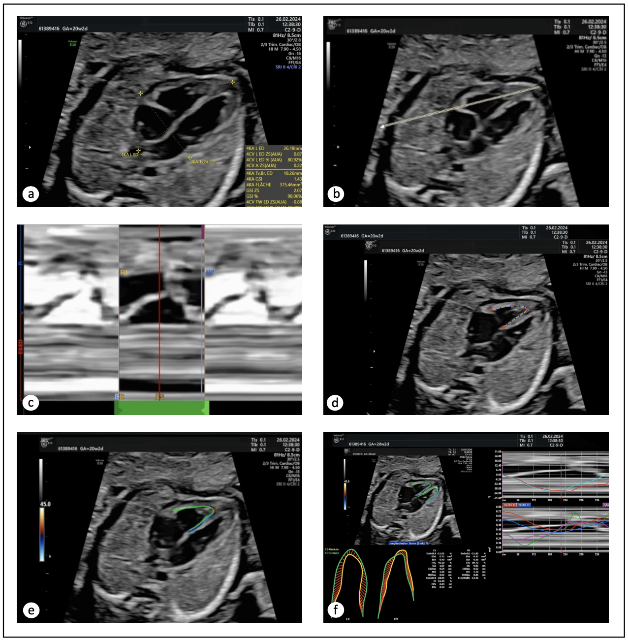
Background Prenatal echocardiographic assessment of fetal cardiac function has become increasingly important. Fetal two-dimensional speckle-tracking echocardiography (2D-STE) allows the determination of global and segmental functional cardiac parameters. Prenatal diagnostics is relying increasingly on artificial intelligence, whose algorithms transform the way clinicians use ultrasound in their daily workflow. The purpose of this study was to demonstrate the feasibility of whether less experienced operators can handle and might benefit from an automated tool of 2D-STE in the clinical routine. Methods A total of 136 unselected, normal, singleton, second- and third-trimester fetuses with normofrequent heart rates were examined by targeted ultrasound. 2D-STE was performed separately by beginner and expert semiautomatically using a GE Voluson E10 (FetalHQ®, GE Healthcare, Chicago, IL, USA). Several fetal cardiac parameters were calculated (end-diastolic diameter (ED), sphericity index (SI), global longitudinal strain (EndoGLS), fractional shortening (FS)) and assigned to gestational age (GA). Bland-Altman plots were used to test agreement between both operators. Results The mean maternal age was 33 years, and the mean maternal body mass index prior to pregnancy was 24.78 kg/m 2. The GA ranged from 16.4 to 32.0 weeks (average 22.9 weeks). Averaged endoGLS value of the beginner was -18.57 % ± 6.59 percentage points (pp) for the right and -19.58 % ± 5.63 pp for the left ventricle, that of the expert -14.33 % ± 4.88 pp and -16.37 % ± 5.42 pp. With increasing GA, right ventricular endoGLS decreased slightly while the left ventricular were almost constant. The statistical analysis for endoGLS showed a Bland-Altman-Bias of -4.24 pp ± 8.06 pp for the right and -3.21 pp ± 7.11 pp for the left ventricle. The Bland-Altman-Bias of the ED in both ventricles in all analyzed segments ranged from -0.49 mm ± 1.54 mm to -0.10 mm ± 1.28 mm, that for FS from -0.33 pp ± 11.82 pp to 3.91 pp ± 15.56 pp and that for SI from -0.38 ± 0.68 to -0.15 ± 0.45. Conclusions Between both operators, our data indicated that 2D-STE analysis showed excellent agreement for cardiac morphometry parameters (ED and SI), and good agreement for cardiac function parameters (EndoGLS and FS). Due to its complexity, the application of fetal 2D-STE remains the domain of scientific-academic perinatal ultrasound and should be placed preferably in the hands of skilled operators. At present, from our perspective, an implementation into clinical practice ‘on-the-fly’ cannot be recommended.