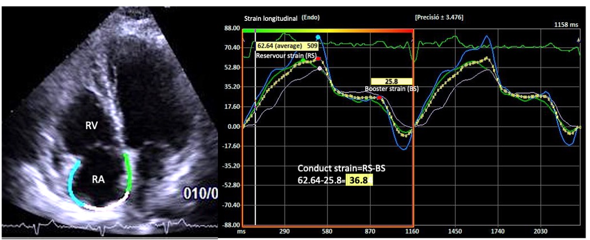
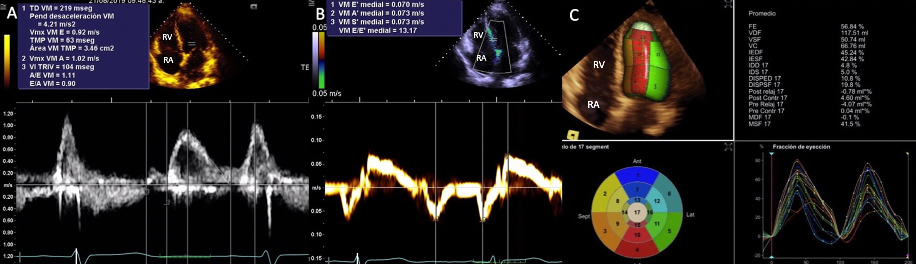
BACKGROUND: Increased systolic pulmonary artery pressure (sPAP) could lead to mechanical dysfunction and myocardial fibrosis of right heart chambers. Echocardiographic strain analysis has not been adequately studied in patients living with pulmonary hypertension (PH). METHODS AND RESULTS: A cross-sectional cohort of patients with suspected PH and echocardiographic strain evaluation was recruited. Cut-off values of peak tricuspid regurgitation velocity with low probability of PH (≤ 2.8 m/s), intermediate probability (2.9-3.4 m/s, without other echo PH signs) and high probability of PH (2.9-3.4 m/s with other echo PH signs and >3.4 m/s) categories were studied by right ventricular and right atrial strain analysis in a sample of 236 patients, 58 (56.9%) had low, 15 (14.7%) intermediate, and 29 (28.4%) high probability of PH. We observed a negative association between right ventricular free wall strain and atrial global strain with sPAP. As PH severity increased, right atrial reservoir, conduit, and contraction (booster) strain values decreased. Identified cut-off values of strain parameters had an adequate ability to detect PH severity categories In addition, post-mortem biopsies of right heart chambers from subjects with known severe pulmonary hypertension were analyzed to quantify myocardial fibrosis. Our sample of right heart biopsies (n=12) demonstrated an association between increased sPAP before death and right ventricular and right atrial fibrosis. CONCLUSIONS: Mechanical dysfunction and fibrosis in right chambers is associated with increased sPAP. Right ventricular and atrial strain could provide enhancement in the diagnosis and categorization of subjects with suspected PH.