Bradley Knight

and 6 more

Background and Aims: The extravascular implantable cardioverter defibrillator (EV ICD) has an extended projected battery longevity compared to the subcutaneous implantable cardioverter defibrillator (S-ICD). This study used modeling to characterize the need for generator changes, long-term complications, and overall costs for both the EV ICD and S-ICD in healthcare systems of various countries . Methods: Battery longevity data were modelled using a Markov model from averages reported in device labelling for the S-ICD and with engineering estimates based on real life usage from EV ICD Pivotal Study patient data to introduce variability. Clinical demographic data of recipients were derived from published literature. The primary outcomes were defined as the number of generator replacement surgeries, complications, and total healthcare system costs due to battery depletion over the expected lifetime of patients receiving EV ICD or S-ICD therapy. A one-way sensitivity analysis of the model was performed for the US healthcare system. Results: Average modelled battery longevity was determined to be 7.3 years for the S-ICD compared to 11.8 years for the EV ICD. The probability of a complication after a replacement procedure was 1.4%, with an operative mortality rate of 0.02%. The use of an EV ICD was associated with 1.4-1.6 fewer replacements on average over an expected patient lifetime as compared to an S-ICD and a 24.3-26.0% reduction in cost. The US sensitivity analysis found use of an EV ICD resulted in a reduction in replacement surgeries of greater than 1 (1.1-1.6) along with 5-figure cost savings in all scenarios ($18,602-$40,948). Conclusion: The longer projected battery life of the EV ICD compared to the S-ICD has the potential to meaningfully reduce long-term morbidity and healthcare resources related to generator changes from the perspective of multiple diverse healthcare systems.

Jeremiah Wasserlauf

and 1 more

Application of electrocautery to a metal guidewire can be used to perform transseptal puncture (TSP). Dedicated radiofrequency guidewires (RF) may represent a better alternative. This study compares safety and effectiveness of electrified guidewires to a dedicated RF wire. TSP was performed on porcine hearts using an electrified 0.014” or 0.032” guidewire under various power settings compared to TSP using a dedicated RF wire with 5W power. The primary endpoint was the number of attempts required to achieve TSP. Secondary endpoints included the rate of TSP failure, TSP consistency, effect of the distance between tip of the guidewire and the tip of the dilator, and effect of RF power output level. Qualitative secondary endpoints included tissue puncture defect appearance, thermal damage to the TSP guidewire or dilator, and tissue temperature using thermal imaging. The RF wire required 1.10 ± 0.47 attempts to cross the septum. The 0.014” electrified guidewire required 2.17 ± 2.36 attempts (2.0x higher than the RF wire; p<0.01), and the 0.032” electrified guidewire required 3.90 ± 2.93 attempts (3.5x higher than the RF wire; p<0.01). Electrified guidewires had a higher rate of TSP failure, larger defects, more tissue charring, higher temperatures, and greater tissue heating. Fewer RF applications were required to achieve TSP using a dedicated RF wire compared to an electrified guidewire. Smaller defects and lower tissue temperatures were also observed using the RF wire. Electrified guidewires required greater energy delivery and were associated with equipment damage and tissue charring.

Rachel Kaplan

and 13 more

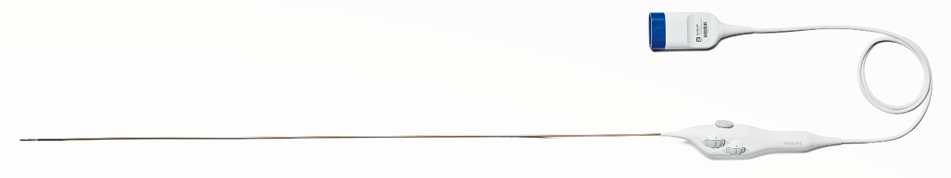
Introduction: Standard two-dimensional (2D), phased-array intracardiac echocardiography (ICE) is routinely used to guide interventional electrophysiology (EP) procedures. A novel four-dimensional (4D) ICE catheter (VeriSight Pro®, Philips, Andover, MA) can obtain 2D and three-dimensional (3D) volumetric images and cine-videos in real time (4D). The purpose of this study was to determine the early feasibility and safety of this 4D ICE catheter during EP procedures. Methods: The 4D ICE catheter was placed from the femoral vein in ten patients into various cardiac chambers to guide EP procedures requiring transseptal catheterization, including ablation for atrial fibrillation and left atrial appendage closure. 2D- and 3D- ICE images were acquired in real time by the electrophysiologist. A dedicated imaging expert performed digital steering to optimize and post-process 4D images. Results: Eight patients underwent pulmonary vein isolation (cryoballoon in 7 patients, pulsed field ablation in 1, additional radiofrequency left atrial ablation in 1). Two patients underwent left atrial appendage closure. High quality images of cardiac structures, transseptal catheterization equipment, guide sheaths, ablation tools, and closure devices were acquired with the ICE catheter tip positioned in the right atrium, left atrium, pulmonary vein, coronary sinus, right ventricle, and pulmonary artery. There were no complications. Conclusion: This is the first-in-human experience of a novel deflectable 4D ICE catheter used to guide EP procedures. 4D ICE imaging in safe and allows for acquisition of high-quality 2D and 3D images in real-time. Further use of 4D ICE will be needed to determine its added value for each EP procedure type.